

Bringing intelligence
to imaging
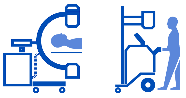
DMS Imaging is focused on the development, design and manufacturing of medical imaging systems mainly for digital radiology and bone densitometry.
Octobre 2023
Nous allons dans le sens d'𝙪𝙣𝙚 𝙖𝙪𝙜𝙢𝙚𝙣𝙩𝙖𝙩𝙞𝙤𝙣 𝙙𝙚𝙨 𝙗𝙚𝙨𝙤𝙞𝙣𝙨 𝙚𝙣 𝙨𝙖𝙣𝙩𝙚́ en raison du 𝙫𝙞𝙚𝙞𝙡𝙡𝙞𝙨𝙨𝙚𝙢𝙚𝙣𝙩 𝙙𝙚 𝙡𝙖 𝙥𝙤𝙥𝙪𝙡𝙖𝙩𝙞𝙤𝙣 et de l'augmentation, avec l'âge, de l'incidence des 𝙥𝙖𝙩𝙝𝙤𝙡𝙤𝙜𝙞𝙚𝙨 𝙘𝙝𝙧𝙤𝙣𝙞𝙦𝙪𝙚𝙨.
21/11/2023

DMS group à l'Elysée dans le cadre de ETIncelles. Programme qui valorise les PME à très fort potentiel.
OUR VISION
DMS is a French industrial company specialized in digital radiology, with an international reach, and recognized as a key actor and an indispensable partner in creating value through the quality of our solutions as well as our flexibility, ingenuity, and socially engaged values.
OUR ENGAGEMENT
ECOSYSTEM
To be a model and a committed unifier of our ecosystem, through the strength of our collaborations with our institutional and industrial partners in France, Europe and around the world.
SHAREHOLDERS
To execute our strategic plan, Imaging 2027, while being attentive to opportunities, to ensure profitable and sustainable growth and the creation of value for the DMS Group and its shareholders, imprinted with environmental and social values.
CUSTOMERS
To be a partner who listens, and who relies on the agility, commitment and competence of our employees to provide customers with differentiating solutions that bring value and offer an optimal quality of service.
COLLABORATORS
To support all employees so that they recognize themselves in the company's values, find pride and meaning in their work, are responsible and know how to take initiatives, and are aware of our shared challenges and have confidence in the collective.
PROJET MC2
Our MC2 project was selected for funding as part of a French government initiative to stimulate the French economy in strategic industrial sectors « AAP Plan de relance pour l’industrie – Secteurs stratégiques ». The funds will allow us to develop and manufacture 2 medical imaging solutions : a mobile x-ray and a surgical C-Arm.